Figure 5.
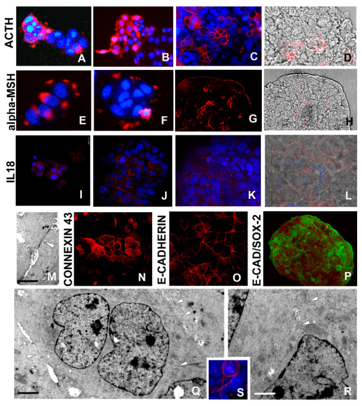
Morpho-functional characterization of cells in developed spheroids: ACTH/α-MSH, interleukin (IL)18 expressions and opening of stable intercellular bridges (A–H). Laser confocal microscope analysis. Representative micrographs depicting ACTH and α-MSH expression by immunocytochemical characterization. At 24 h (A,E) and 3 days (B,F) spheroid culture, the signal is quite strong, whereas in mature spheroids (C,D,G,H), the signal is superficially localized. In D and H panels, red signal (immunolocalization) and bright field were superimposed to better identify the involved area of spheroid. (I–L) IL18 expression is localized in most cells in early aggregates (I,J) and especially in peripheral cells of mature spheroid (K,L). In L panel, red signal (immunolocalization) and bright field were superimposed. (M) TEM analysis. Two neighboring cells are in close contact according to the presence of specialized junctions (arrowhead). Scale bar: (M) 2 μm. (N–P) Confocal microscopy images. Starting from 5–7 days, cells are connected by gap junctions that are characterized by Connexin 43 (N) and E-cadherin (O) expression. Staining with antibody raised against E-cadherin (O) shows a punctate pattern at the periphery of the cells. (P) Confocal microscopy images. Double labelling of spheroid cells with antibodies raised against E-cadherin (red) and Sox2 (green) showing that stemness (Sox2) does not match with the gap formation that is considered critical event in growth and differentiation. (Q,R) TEM analysis of MCF7 cells showing the presence of intercellular bridges that allow cytoplasmic continuity (arrowheads). Scale bars: (Q) 2.5 μm: (R) 2 μm. (S) Immunofluorescence staining showing expression of TEX14 (red signal) lining the MCF7 cells. Nuclei are stained with DAPI.

