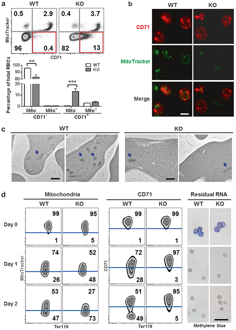
Fig. 2.

Loss of mitochondria in IEX-1 KO reticulocytes while retaining CD71 on the cells. a–c IEX-1 KO CD71+ reticulocytes were absent of mitochondria. Peripheral blood cells of WT and KO mice were stained with Ter119, CD71, and MitoTracker, counted by flow cytometry (upper) and statistically analyzed (lower) (a). IEX-1 KO mice had an abnormal population of CD71+Mito− cells highlighted in red, within Ter119+ RBCs. Data were presented as mean ± SEM (n = 12), **P < 0.01 and ***P < 0.001 compared between WT and KO cells. Ter119+CD71+ reticulocytes were sorted from WT and KO peripheral blood cells for analyzing mitochondrial content by confocal microscopy (b; scale bar, 5 μm) or transmission electron microscopy (c; mitochondria, blue arrows; scale bar, 500 nm). d Accelerated mitochondrial clearance and delayed CD71 shedding in KO reticulocytes during maturation. Ter119+ CD71high reticulocytes were sorted from WT and KO mice 3 days after PHZ treatment and cultured for 2 days. MitoTracker, anti-CD71 antibody, and Methylene blue were then used to stain mitochondria, CD71, and residual RNA, respectively. Scale bar, 20 μm.
