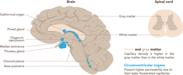
Fig. 4.
Regional differences in the brain microvasculature. Schematic representation of a brain sagittal section (left) and a spinal cord transverse section (right). Capillary density is higher in the CNS gray matter than in the white matter, according to their differential metabolic activity. The white matter of the corpus callosum is highlighted. The microvessels in the circumventricular organs (CVOs, highlighted in blue) lack BBB characteristics, rather they are fenestrated and thus permeable to blood components. CVOs include the subfornical organ, the vascular organ of the lamina terminalis, the area postrema, the median eminence, the neurohypophysis, the pineal gland, and the choroid plexus

