Fig. 4.
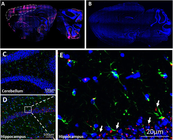
MSC-exo can cross the BBB and integrate into the cells in the tissue. a Complete sagittal section of Shank3B KO shows MSC-exo are found in the parenchyma and accumulate mainly in the area of the cortex, cerebellum, and some accumulation in the hippocampus (96 h post intranasal administration). b Complete sagittal section of WT shows complete evacuation of MSC-exo from the brain (96 h post intranasal administration). c, d Magnification of the cerebellum and hippocampus tissues of Shank3B KO shows MSC-exo are found in the tissue. e Magnification of the CA1 area with DAPI (blue), PKH26 exosomes (red), and astrocytes (GFAP green)

