Fig. 6.
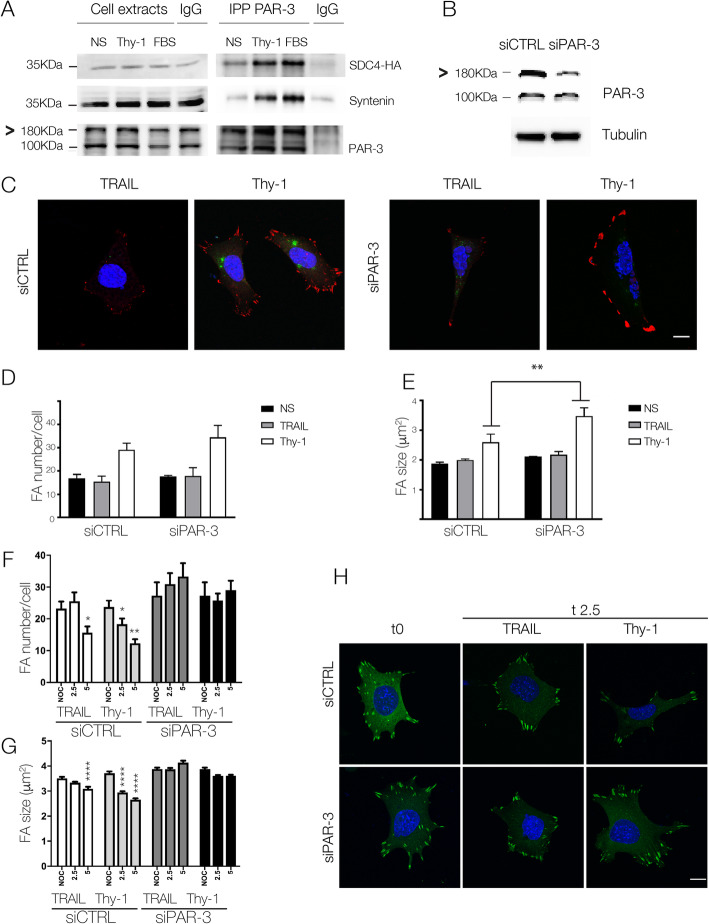
Thy-1-induced Syndecan-4/PAR-3 association is needed in FA dynamics in mouse embryonic fibroblasts. a MEFs transfected with Syndecan-4-HA were stimulated or not (NS) with Thy-1-Fc or fetal bovine serum (FBS). PAR-3 was immunoprecipitated (IP) from the lysates, and Syndecan-4 was detected with an anti-HA antibody (SDC4-HA). Syntenin and PAR-3 were immunoblotted as positive and an IP control, respectively. IgG was used as a negative IP control. b MEFs transfected with the siRNA control or PAR-3 were analyzed by Western blotting for PAR-3 expression. Tubulin was used as a loading control. c MEFs co-transfected with siGlo and siRNA control or PAR-3 were stimulated with Thy-1-Fc-Protein A (Thy-1) or TRAIL-R2-Fc-Protein A (TRAIL) for 15 min. FAs and nuclei were respectively visualized by vinculin staining (red) and DAPI (blue). Transfected cells were identified by the siGlo signal (green). Graphs show the mean ± s.e.m. for the number (d) and average area (e) of FAs from at least 50 cells. MEFs were transfected as in (c), incubated with Nocodazole and washout, and immediately stimulated as in (c) for different time periods. Graphs show the mean ± s.e.m. for the number (f) and average area (g) of FAs from at least 50 cells. Significant differences are indicated as *P < 0.05, **P < 0.01, and ***P < 0.001. (h) Cells were co-transfected with Paxillin-EGFP and siRNA (control or PAR-3). Shown are representative images for non-stimulated cells (t0) and cells stimulated with Thy-1-Fc-Protein A (Thy-1) or TRAIL-R2-Fc-Protein A (TRAIL) for 2.5 min (t2.5). Scale bar =10 μm

