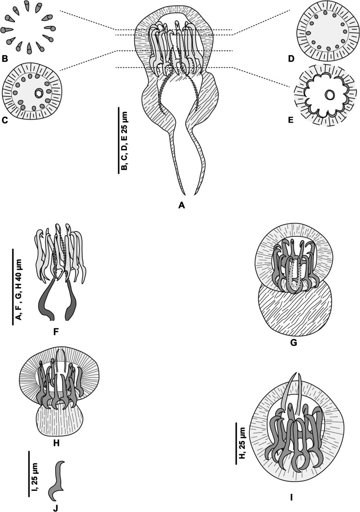
Figure 3.
Pseudaxine trachuri, genital atrium shown laterally and in different optical sections and the variable position of the stylet. (A) Genital atrium, lateral view, MNHN HEL1284; (B–E), optical sections of a genital atrium in perfect apical view; the level of these sections are shown on the lateral view, MNHN HEL1285. (F) Lateral view of the atrium showing only the sclerotised parts, MNHN 306HG, Box 43, slide 51; (G–I) various views at different orientations, MNHN HEL1286, MNHN HEL1287, MNHN HEL1288. (J) One spine, MNHN HEL1287.

