. 2020 Aug 14;35(7):e202000701. doi: 10.1590/s0102-865020200070000001

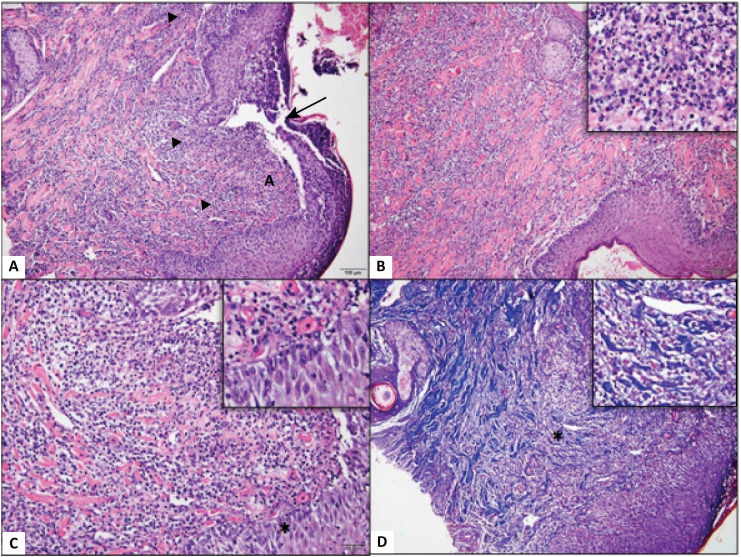
Figure 2. Photomicrograph of dog skin. (A) Skin with solution of continuity of the epidermis (arrow), associated with a marked neutrophilic inflammatory infiltrate (arrowhead) diffusely distributed through the dermis. (B) Note that the intense neutrophilic infiltrate (Detail, x20) is widely distributed in the superficial dermis. (C) Observe moderate edema of the epidermis evidenced by the increase in the space between the keratinocytes (*, Detail, x40), in addition to the marked inflammatory infiltrate. Hematoxylin and Eosin (x10). (D) Dissociation of collagen fibers from the dermis associated with intense and diffuse inflammatory infiltrate in the dermis (*, Detail, x20). Masson's trichrome, Obj. x10.

Figure 2