
Figure 5. Topographic Restriction of the Striatonigral Projection According to the Resident Sector in the Striatum.
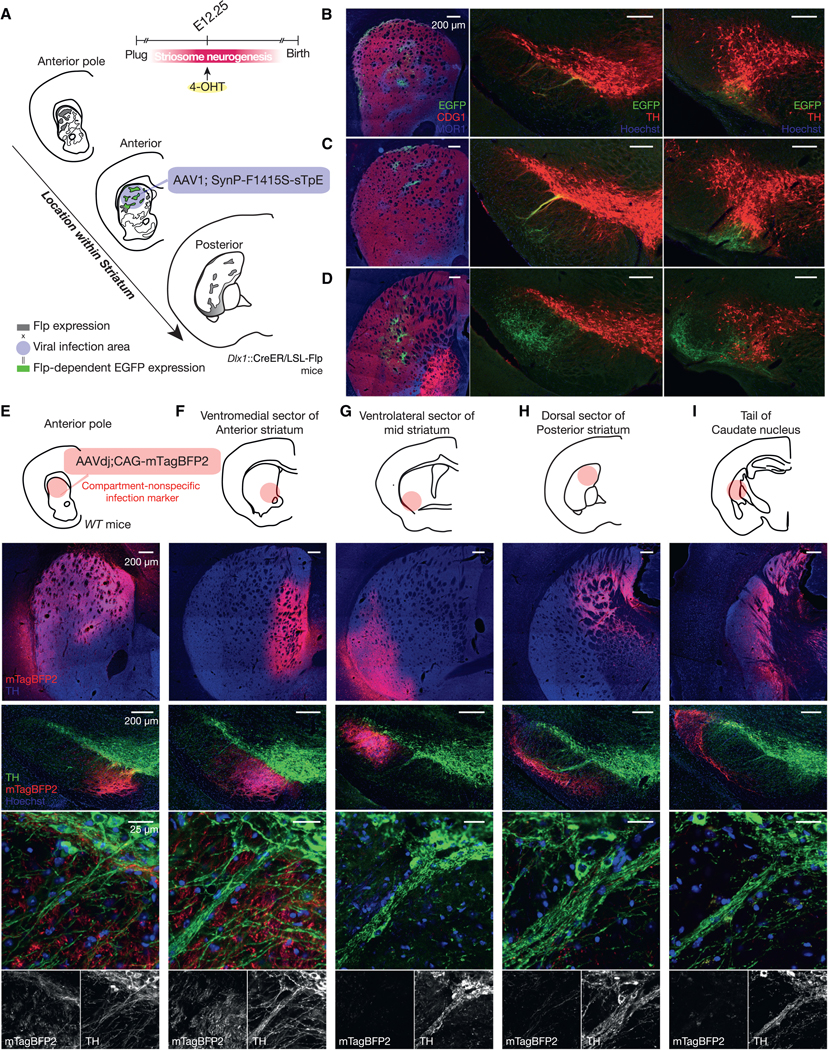
(A) Anterograde tracing of striatonigral axons from same-age SPNs settled in different functional sectors.
(B) Striatonigral projections of E12.25-born SPNs settled in the anterior pole of striatum. Left: striatal injection site stained for MOR1 (blue), CDG1 (red), and EGFP(green). Middle and right: anterior (middle) and posterior (right) SN stained for TH (red), GFP (green), and Hoechst (blue). Number of mice = 3.
(C and D) Same as in (B) but for E12.25-born SPNs settled in the anterior (C) and posterior (D) striatum. Number of mice = 3.
(E–I) Compartment-nonspecific infection marker was injected into the anterior pole (E), ventromedial/limbic sector of anterior striatum (F), ventrolateral sector of mid striatum (G), dorsal sector of posterior striatum (H), or tail of caudate nucleus (I). Upper panels: injection sites stained for mTagBFP2 (red) and TH (blue). Lower panels: nigral sections of the same mice stained for TH (green), mTagBFP2 (red), and Hoechst (blue). None of them route axons of resident SPNs to the central striosome-dendron bouquet. Number of mice = 2.6 ± 0.55.