
Fig. 10—
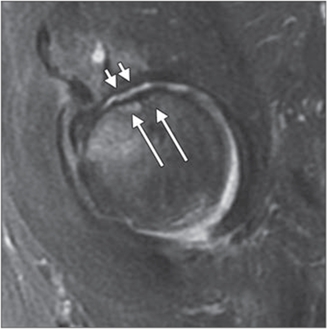
Sagittal intermediate-weighted fat-suppressed fast spin-echo image of 65-year-old woman shows femoral head subchondral insufficiency fracture (long arrows) and joint space narrowing. Diffuse cartilage lossisseen in weight-bearing central and anterior regions of acetabulum and femoral head (short arrows). There is also mild bone marrow edema pattern in femoral head adjacent to fracture.