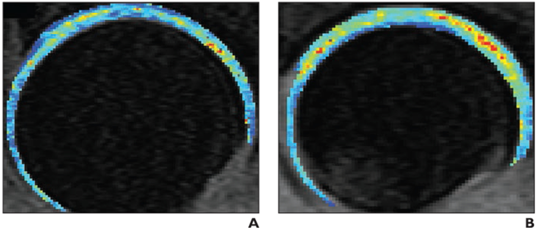
Fig. 3—

T1rho color maps of acetabular cartilage and femoral cartilage. (Courtesy of Samaan M and Souza RB, University of California at San Francisco, San Francisco, CA)
A and B, Composite T1rho color maps of both acetabular cartilage and femoral cartilage of left hip obtained in sagittal plane in healthy 35-year-old control subject (A) and 30-year-old woman with early hip osteoarthritis (B). T1rho values are elevated (yellow, orange, and red) in weight-bearing regions of osteoarthritis patient (in particular, in superolateral and superomedial acetabular and femoral cartilage), indicating changes in regional proteoglycan composition. Also note that patient (B) does not have joint space narrowing and that joint space appears mildly widened compared with control subject (A); joint space widening in patient potentially reflects cartilage swelling. Blue and green = normal T1rho values.
