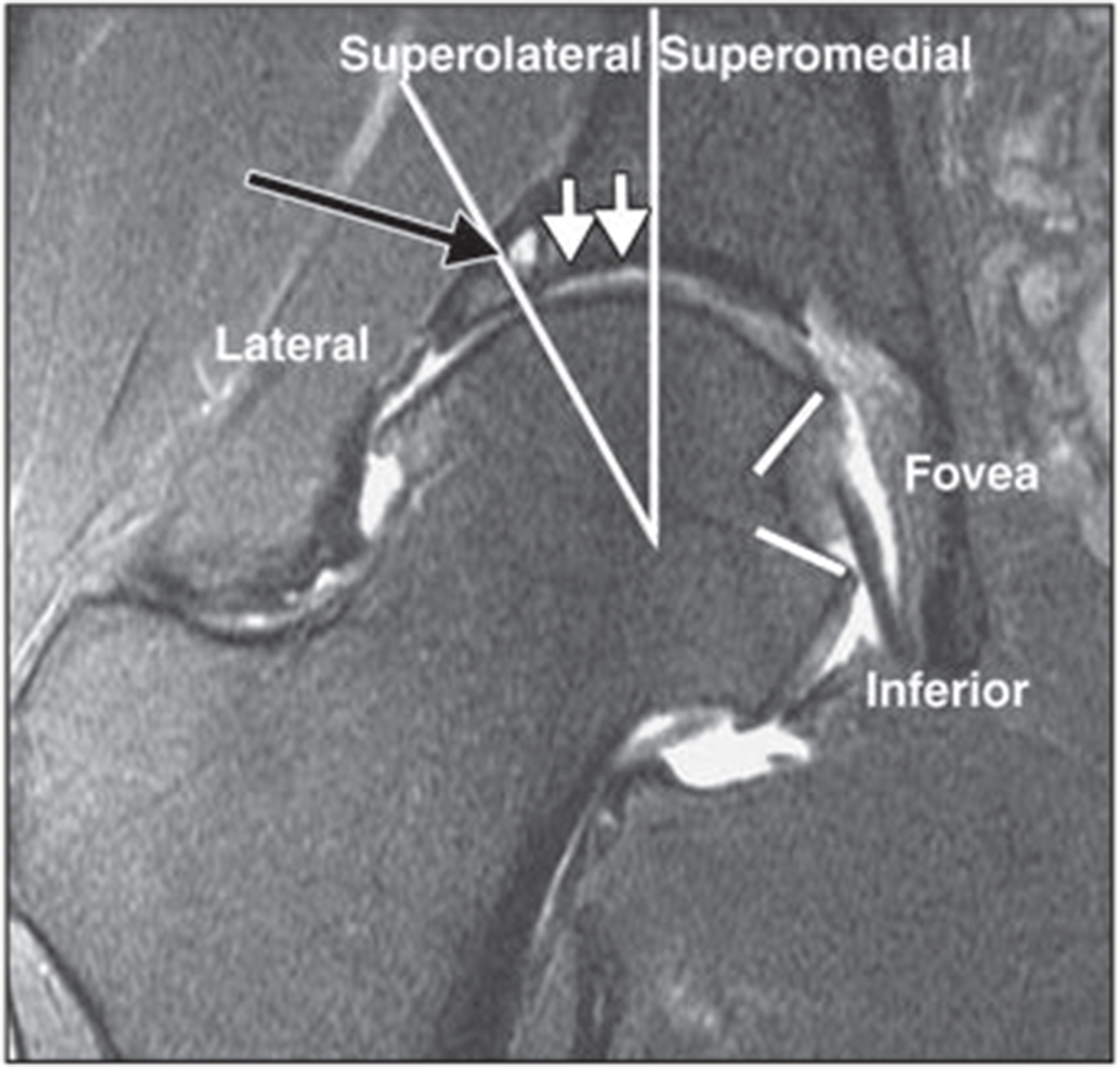
Fig. 4—

Coronal 2D fat-suppressed intermediate-weighted fast spin-echo image of right hip of 42-year-old woman with degenerative disease of hip and femoroacetabular impingement. According to score obtained using SHOMRI (scoring hip osteoarthritis with MRI) scoring system [21], femoral cartilage is separated into four subregions and acetabular cartilage into three subregions in coronal image. There is full-thickness acetabular cartilage loss with diameter of 10 mm (white arrows) in superolateral subregion, which would be scored as grade 2 lesion according to SHOMRI. There is also subchondral cyst (black arrow) in superolateral region. Findings were arthroscopically confirmed. Long lines show separate lateral, superolateral, and superomedial regions, and short lines = indicate foveal regions.
