Fig. 6—

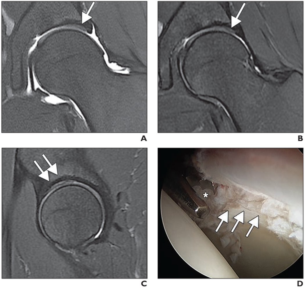
MR arthrography of left hip of 26-year-old man with clinical diagnosis of femoroacetabular impingement.
A and B, Unenhanced coronal intermediate-weighted fat-saturated fast spin-echo (FSE) MR arthrograms obtained at 1.5 T (A) and at 1.5 T, unehanced 3-T MRI, and arthroscopy at 3T (B) show area of bright signal intensity along superior acetabulum with adjacent darker hypointense cartilage (arrow) consistent with delamination.
C, Sagittal intermediate-weighted fat-saturated FSE image obtained at 3 T shows findings (arrows) similar to coronal images (A and B).
D, Arthroscopic image shows acetabular cartilage delamination (arrows) with injured cartilage layer being débrided by arthroscopic biter (asterisk).
