
Fig. 7—
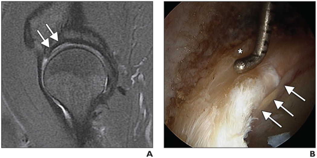
Cartilage delamination in 55-year-old man with clinical signs of femoroacetabular impingement.
A, Sagittal intermediate-weighted fat-saturated fast spin-echo (FSE) image obtained at 3 T shows abnormal cartilage at acetabulum with bright signal intensity along subchondral bone and layer of darker hypointense cartilage covering this area (arrows).
B, Arthroscopic image shows acetabular cartilage delamination at chondrolabral junction as pressure applied to labrum by arthroscopic probe (asterisk) causes positive wave sign to underlying delaminated cartilage (arrows).