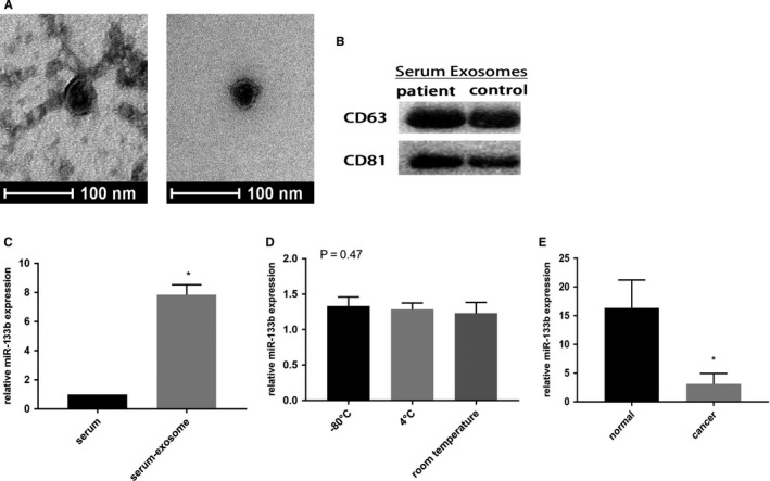
Figure 2.

Expression of serum exosomal miR‐133b in patients with bladder cancer. Transmission electron microscopy image of exosomes derived from the serum of patients and controls. Scale bars represent 100 nm (A). Western blotting analysis showing the presence of CD63 and CD81 in exosomes (B). The expression of miR‐133b was detected in serum and serum exosomes (C). The relative expression levels of exosomal miR‐133b were stable after storing at −80°C, 4°C and room temperature for 12 hours respectively (D). qRT‐PCR detection of miR‐133b in exosomes from serum (E). *P < .05
