Figure 1.

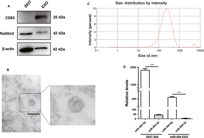
Characteristics of exosomes derived from miR‐204‐5p‐overexpressing HEK293T cells. A, The markers of exosomes (CD63 and Flotillin‐2) were detected in HEK293T cells and exosomes by Western blot. B, The transmission electron micrograph showed roundshaped vesicles with bilayered membranes ranging from 100 nm to 150 nm in diameter released by HEK293T cells. Scale bar = 200 nm. C, 293T EXOs size distribution was measured by Zetasizer. D, Real‐time qRT‐PCR revealed that the level of miR‐204‐5p was higher in 293T‐204 cells and miR‐204 EXO than miR‐204‐3p. ***P < .001. Shown are mean ± SEM from three independent experiments
