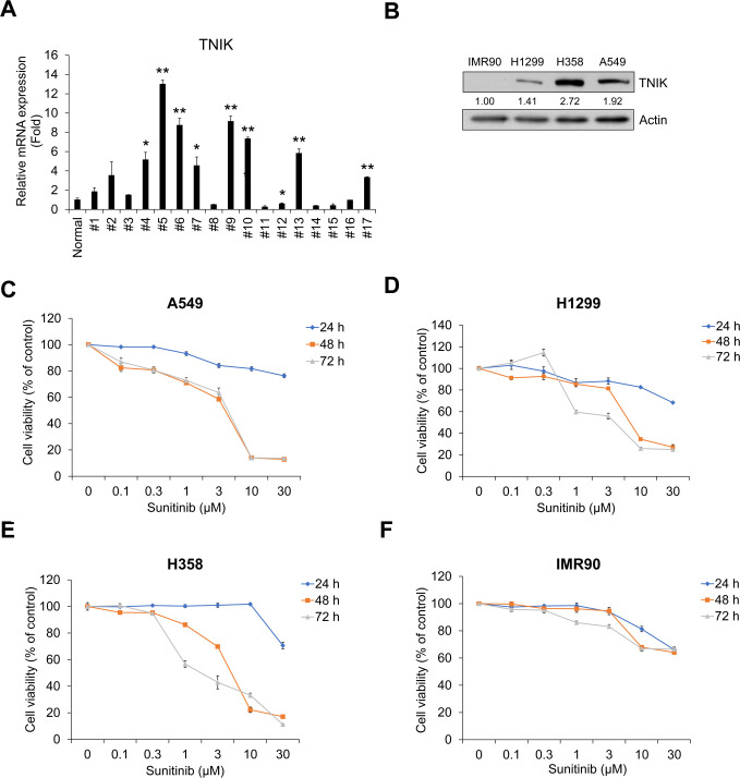
Fig 2. Sunitinib inhibits the proliferation of TNIK-expressing human NSCLC cells.
(A) TNIK mRNA expression in NSCLC tissue cells from lung cancer patients. TNIK mRNA expression levels were measured by qRT-PCR analysis in pool normal lung tissue cells (n = 8) and in NSCLC cells from patients with lung cancer (n = 17). The normal group represents the average qRT-PCR results obtained from eight healthy lung tissue cells without cancer cells. Data represent the mean ± SD of raw results. Experiments were performed in triplicate. * p < 0.05 and ** p < 0.001 (vs. control). (B) The protein expression of TNIK in normal fibroblast IMR90 and NSCLC H1299, H358, and A549 cells was measured by Western blot. Actin was used as a loading control. A549 (C), H1299 (D), and H358 (E) cells and IMR90 (F) cells were treated with sunitinib for 24‒72 h. After incubation, cell viability was measured by CCK-8 assay. Experiments were performed in triplicate. Data represent the mean ± SD of raw results.

