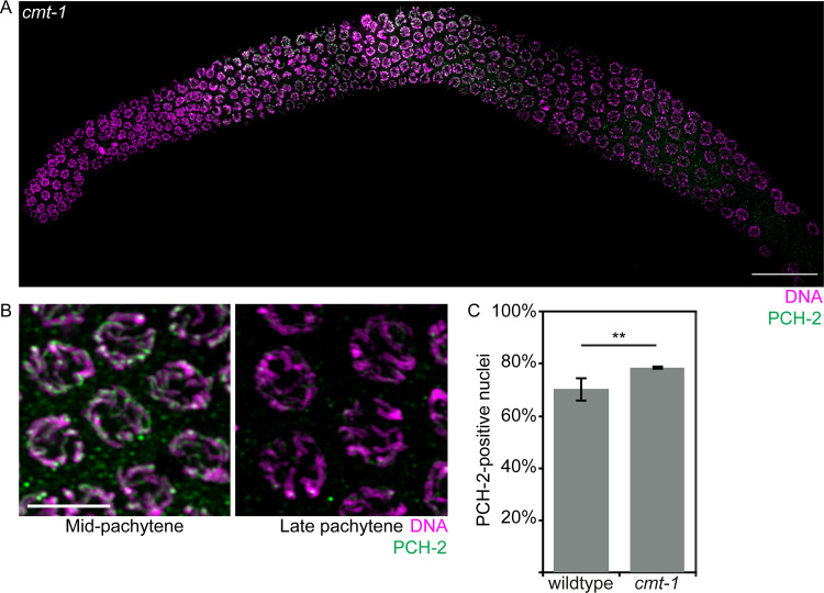
Fig 7. PCH-2 localizes to meiotic chromosomes in cmt-1 mutants.
A. Whole germline image of PCH-2 and DAPI staining in a cmt-1 mutant germline. Scale bar indicates 20 microns. B. Meiotic nuclei in mid-pachytene stained with DAPI and antibodies against PCH-2 in cmt-1 mutants. C. Quantification of percentage of PCH-2-positive nuclei in wildtype and cmt-1 mutant germlines. Error bars indicate 95% confidence intervals. Significance was assessed by performing two-tailed t-tests. A ** indicates a p value < 0.01.

