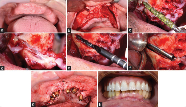
Figure 5.
Workflow of zygomatic implant insertion. (a) Deficient maxilla. (b) Mucoperiosteal flap. (c) Tunnel formation through the residual alveolar bone from the center of the ridge. (d) Elongated window lateral to the maxillary sinus and reflection of the Schneiderian membrane. (e) Marking the point of entry to the zygomatic bone and drilling into the zygoma. (f) The zygomatic implant is inserted in a high torque. Only the apical one third of the implant is inserted into the bone. (g) Angulated multi-unit abutments were used to correct the angulations. (h) Immediate prosthetic rehabilitation

