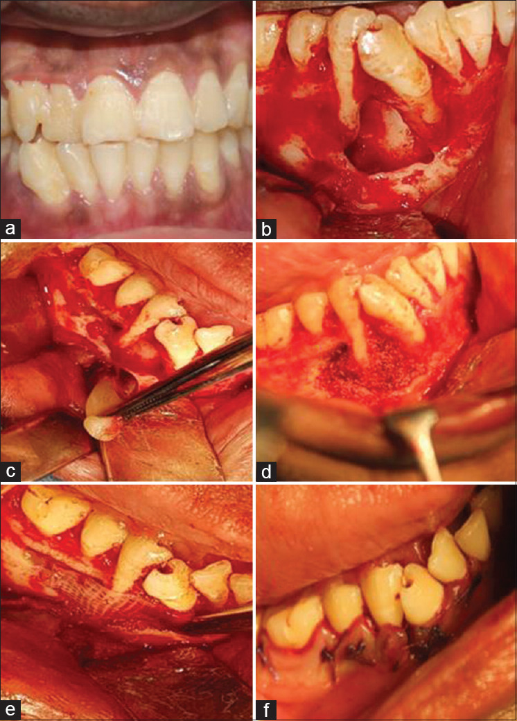
Figure 1.

Preoperative clinical and surgical procedure. Preoperative clinical photograph showing drifted 43, 44, 45 (a). Flap elevation and cystic enucleation, apicoectomy, and retrograde filling (b). Placement of concentrated growth factor cystic space (c). Placement of sticky bone (d). Placement of concentrated growth factor membrane (e). Sutures placed (f)
