Image 2.

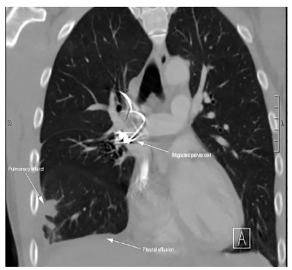
Computed tomography imaging showing a migrated pelvic coil (mid-image arrow) in the right pulmonary artery with areas of pulmonary infarct in the right middle and lower lobes and a small right pleural effusion (left and lower arrows).

Computed tomography imaging showing a migrated pelvic coil (mid-image arrow) in the right pulmonary artery with areas of pulmonary infarct in the right middle and lower lobes and a small right pleural effusion (left and lower arrows).