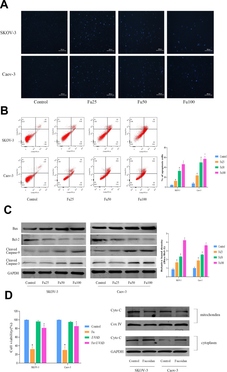
Figure 3.
The effects of fucoidan on OC cells apoptosis. (A) Nuclear fragmentation of SKOV-3 and Caov-3 cells was observed by fluorescence microscopy and staining with Hoechst 33,342 dye (magnification, 200 ×). (B) Apoptosis of SKOV-3 and Caov-3 cells was determined by flow cytometry. (C) Western blot analysis was performed to determine the levels of proteins related to cell apoptosis. The data are expressed as the mean ± SD (n=3, *p < 0.05 for Fu25 vs Control; #p < 0.05 for Fu50 vs Fu25; and ^p < 0.05 for Fu100 vs Fu50). (D) SKOV-3 and Caov-3 cells were pretreated with or without 50 μM of the pan-caspase inhibitor Z-VAD-FMK for 1 h. Western blot analysis was performed to determine the levels of Cyto C of mitochondria and cytoplasm. The data are expressed as the mean ± SD (n=3, *p < 0.05 for Fu100 vs Fu100+Z-VAD-FMK, ^p < 0.05 for Z-VAD-FMK vs Fu100+Z-VAD-FMK).

