. 2020 Aug 18;9:e58057. doi: 10.7554/eLife.58057

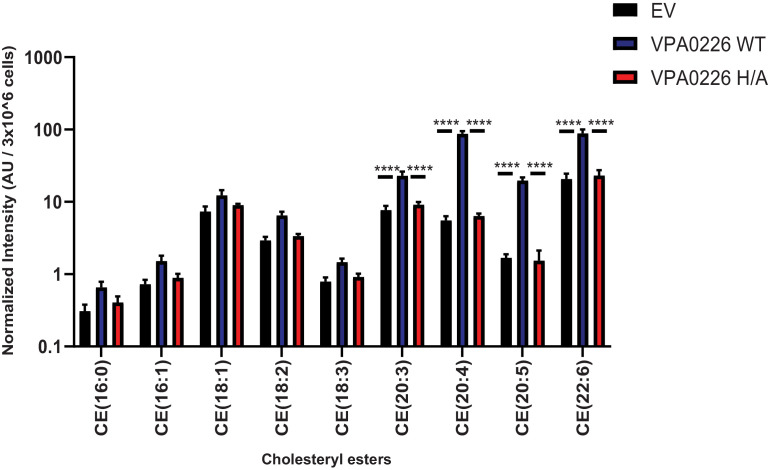
Figure 4. VPA0226 changes lipid profile in cells.

Quantification of polyunsaturated and saturated fatty acyl cholesteryl esters extracted from transfected HeLa cells (empty vector (EV), wildtype VPA0226-sfGFPN1 and VPA0226-H/A-sfGFPN1). Numbers are expressed as an average of five technical replicates for one of three independent experiments. Error bars represent standard deviation from the mean. Asterisks represent statistical significance (****=p < 0.0001) using one-way ANOVA and Turkey’s multiple comparison test.

Figure 4.

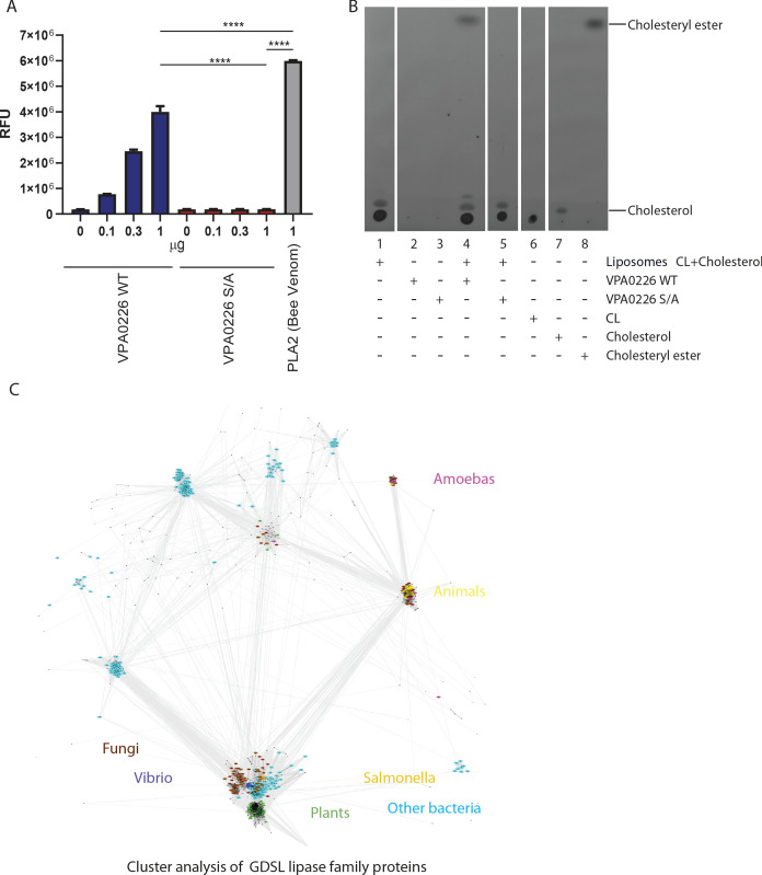
Figure 4—figure supplement 1. VPA0226 displays phospholipase 2 (PLA 2) activity.

Figure 4—figure supplement 1.

(A) Increasing amounts of purified VPA0226WT or catalytically inactive S/A version were incubated with the fluorogenic PLA2-specific substrate BODIPY PC-A2 (EnzCheck Phospholipase A2 Assay kit, Invitrogen) for 30 min at RT and fluorescence was measured at Ex/Em 460/515 nm. Error bars represent standard deviation from the mean. Asterisks represent statistical significance (*=p < 0.05, ***=p < 0.0005) using one-way ANOVA and Turkey’s multiple comparison test. (B) Thin layer chromatography of lipids extracted from the liposome assay demonstrating the formation of esterified cholesterol in reactions containing cardiolipin (CL) and cholesterol liposomes incubated with wildtype VPA0226 (lane 4). For standards, 10 µg each of CL, cholesterol and cholesteryl ester were loaded on TLC plate (lanes 6, 7 and 8, respectively). (C) CLANS graph depicts sequence similarity relationships between GDSL lipases, by clustering protein sequences according to pairwise BLAST-derived scores. 1500 representative sequences of representatives of the Lipase_GDSL family shown. Coloring by taxonomy: Vibrio – dark blue, Salmonella – orange, other bacteria – cyan, fungi – brown, plants – green, animals – yellow, amoebas – magenta.
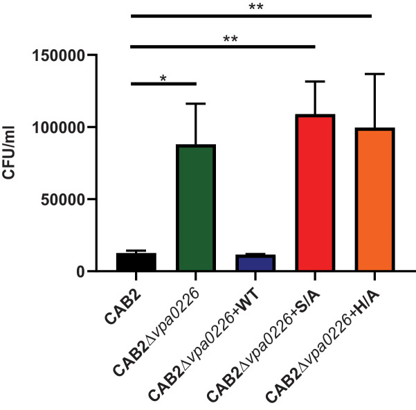
Figure 4—figure supplement 2. CAB2Δvpa0226+H/A mutant displays an egression defect similar to CAB2Δvpa0226 and CAB2Δvpa0226+S/A.

Figure 4—figure supplement 2.

HeLa cells were infected with either CAB2, CAB2Δvpa0226, CAB2Δvpa0226+WT, CAB2Δvpa0226+S/A or CAB2Δvpa0226+H/A for 7 hr PGT. Host cell lysates were serially diluted and plated onto MMM agar plates for intracellular bacterial colony counting (CFU/mL). Numbers are expressed as an average of three technical replicates for one of three independent experiments. Error bars represent standard deviation from the mean. Asterisks represent statistical significance (*=p < 0.05, **=p < 0.01) using 2way ANOVA and Turkey’s multiple comparison test.