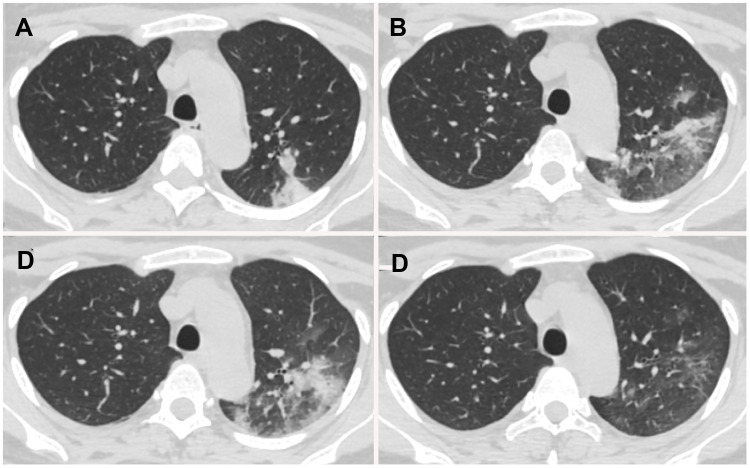
Figure 5.
Chest CT images of a 29-year-old perinatal female with COVID-19 pneumonia, presenting post-partum fever for 1 day. (A) Day 1 after symptom onset, pure consolidation was located peripherally in the left upper lobe. (B) Day 7, the lesion progressed with increased extent and heterogeneous density. (C) Day 11, the lesion further progressed with increased consolidation component. (D) Day 18, lesions were dissipated into ground-glass opacity and few irregular linear opacities.

