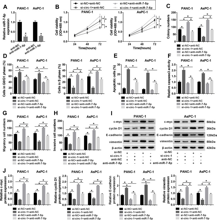
Figure 6.
The effect of miR-7-5p and circ_0013912 in PDAC cells in vitro. (A) RT-qPCR detected miR-7-5p expression in PANC-1 and AsPC-1 cells transfected with miR-7-5p inhibitor (anti-miR-7-5p) or the negative control anti-NC. (B–J) PANC-1 and AsPC-1 cells were co-transfected with si-NC and anti-NC, si-circ-1 and anti-NC, si-NC and anti-miR-7-5p, si-circ-1 and anti-miR-7-5p. (B) CCK-8 assay evaluated cell viability. (C) Colony numbers were determined by colony formation assay. (D, E) FACS method determined the percentages of (D) cells in G0/G1 and S phases, and (E) apoptotic cells. (F) Caspase 3 assay kit estimated caspase 3 activity. (G, H) Transwell assay showed numbers of migratory cells and invasive cells (100×). (I, J) Western blotting examined protein expression of c-myc, cyclin D1, E-cadherin, and vimentin, compared to β-actin. *P<0.05.

