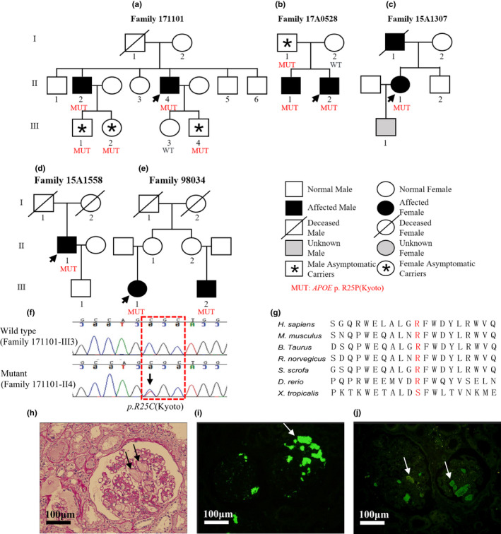
FIGURE 1.

Family pedigrees, genomic sequencing, and kidney biopsy specimens of patients with APOE‐Kyoto mutation. (a–e) Family pedigrees of five lipoprotein glomerulopathy (LPG) families with APOE‐Kyoto mutation. Arrows show the probands. (f) Representative chromatograms of control (upper) and mutant (lower). The position of APOE‐Kyoto mutation is marked by arrow and red dashed box shows the genetic code of arginine (Arg). (g) Multiple sequence alignment of different species shows a high level of conservation at the position of substitution. (h) A representative glomerulus from patient F171101‐II4 shows marked dilatation of the capillary lumen in the glomeruli by a pale‐stained thrombus‐like substance (periodic acid‐Schiff stain). Immunofluorescence shows that apoE (i) and apoB (j) are present primarily in the capillary lumen. GenBank accession no. NM_000041.4. Scale bars 100 μm
