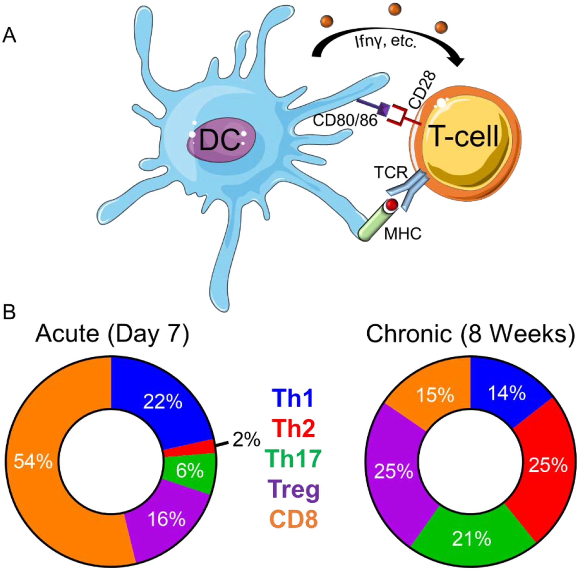
Figure 1: The post-MI myocardial environment facilitates in T-cell activation and phenotype.

A) Antigen-presenting cells such as dendritic cells (DC) activate T-cells via presentation of cognate peptides expressed on major histocompatibility complex (MHC) to T-cell receptors (TCR), surface expression of co-stimulatory signals (i.e. CD28, CD80, CD86), and production of cytokines (IFNγ, etc.). B) T-cell populations change over time. In the acute post-MI environment Th1 and CD8+ are the predominant T-cells whereby in the chronic post-MI environment Th2, Th17, and Treg become the predominant phenotype. The circle charts reflect averages of T-cell populations (%) reported at acute (7 days) and chronic (8 weeks) post-MI remodeling time points.[14, 19–21]
