
Figure 7.
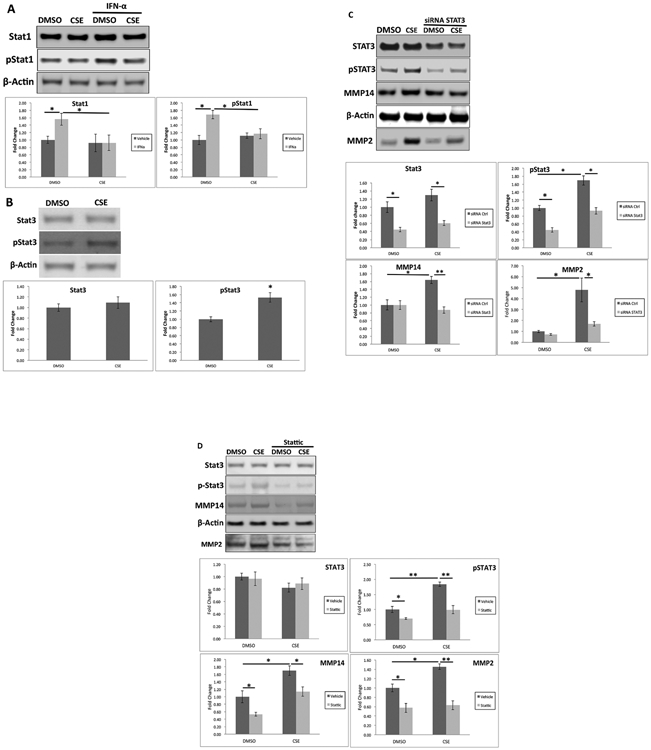
STAT1 is decreased by cigarette smoke extract (CSE). A. RPE cells were treated with DMSO or 125 μg/ml CSE in the presence of 100 ng/ml IFN- α for 6 hrs. Western blots of STAT1 and pSTAT1 and their abundances were plotted as fold change. B. Cells were treated with DMSO or CSE for 24 hrs. Western blots of STAT3 and pSTAT3, and their abundances were plotted as fold change. *p<0.05. STAT3 silencing abrogates CSE-induced MMP14 production and MMP2 secretion. C. ARPE-19 cells were transfected with STAT3 siRNA and treated with 125 μg/ml CSE for 24 h. Western blot shows total STAT3, p-STAT3, and MMP14, and their abundances were plotted as fold change relative to DMSO-treated control siRNA treated cells. Data were normalized to β-actin. D. RPE cells were transfected with STAT3 siRNA, and treated with 125 μg/ml CSE for 24 h. Western blot shows total STAT3, p-STAT3, and MMP14 from cell lysates and MMP2 from the supernatant, and their abundances were plotted as fold change relative to DMSO-treated control siRNA treated cells. Data were normalized to β-actin. *p<0.05; **p<0.01.