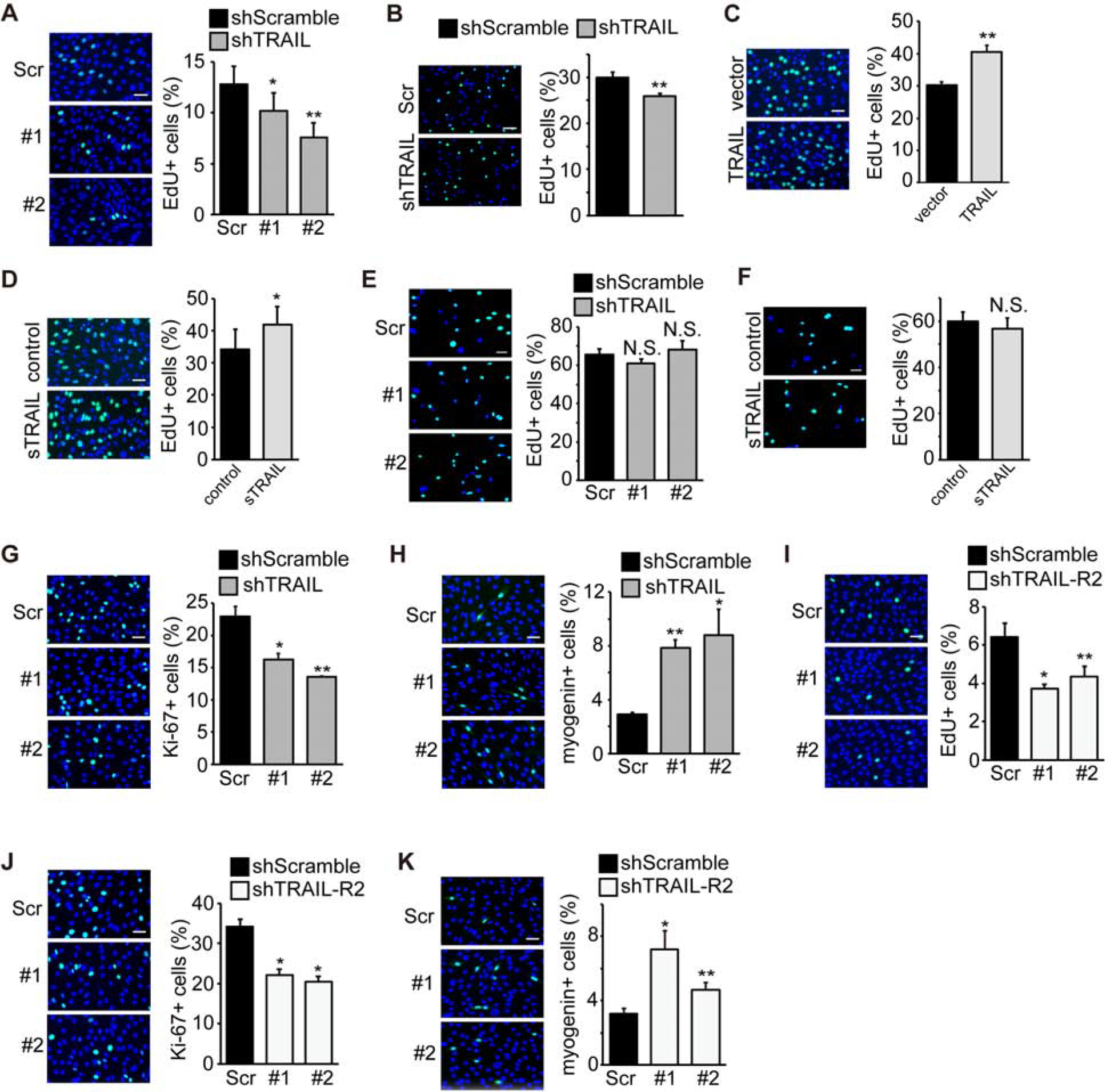
Figure 4. TRAIL and TRAIL-R2 promote myoblast proliferation at the onset of differentiation.

(A) C2C12 cells were transduced with lentiviruses expressing shTRAIL or shScramble followed by 2 days of puromycin selection. At 24 hr of differentiation, cells were labeled with EdU for 2 hrs in differentiation medium, stained for EdU (green) and DAPI (blue), and the percentage of cells positive for EdU staining was quantified (n=6). (B) Mouse primary myoblasts were transduced with lentiviruses expressing shRNAs overnight, cultured in growth medium for 1 day, and then labeled with EdU for 2 hrs in differentiation medium at 0 hr of differentiation and processed as in (A) (n=5). (C) C2C12 cells were transfected with TRAIL or empty vector and selected with hygromycin for 2 days. At 0 hr of differentiation, cells were labeled with EdU for 2 hrs in differentiation medium, and processed as in (A) (n=5). (D) C2C12 cells were grown in the presence of vehicle (Control) or 100 ng/mL sTRAIL for 24 hrs. At 0 hr of differentiation, cells were labeled with EdU for 2 hrs in differentiation medium, and processed as in (A) (n=6). (E) C2C12 cells were transduced with lentiviruses expressing shTRAIL or shScramble followed by 2 days of puromycin selection. Cells were then grown to ~20% confluence, labeled with EdU for 2 hrs in growth medium, and processed as in (A) (n=5). (F) C2C12 cells were grown to ~20% in the presence of vehicle (Control) or 100 ng/mL sTRAIL for 24 hrs, labeled with EdU for 2 hrs in growth medium, and processed as in (A) (n=4). (G) C2C12 cells treated as in (A) were stained for Ki-67 (green) and DAPI (blue), and the percentage of cells positive for Ki-67 staining was quantified (n=4). (H) C2C12 cells treated as in (A) were stained for myogenin (green) and DAPI (blue), and the percentage of cells positive for myogenin staining was quantified (n=4). (I) C2C12 cells were transduced with lentiviruses expressing shTRAIL-R2 or shScramble followed by 2 days of puromycin selection. At 24 hr of differentiation, cells were labeled with EdU for 2 hrs in differentiation medium and processed as in (A) (n=5). (J) C2C12 cells treated as in (I) were stained for Ki-67 (green) and DAPI (blue), and the percentage of cells positive for Ki-67 staining was quantified (n=4). (K) C2C12 cells treated as in (I) were stained for myogenin (green) and DAPI (blue), and the percentage of cells positive for myogenin staining was quantified (n=4). All error bars represent SEM. Two-tailed t-test was performed to compare each data to control. *P < 0.05; **P < 0.01. NS, not significant. Scale bars: 50 μm.
