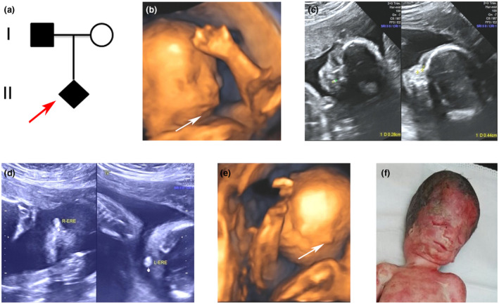
FIGURE 1.

Family pedigree and male fetus at 24 weeks of gestation with craniofacial features by 2D and 3D ultrasound inspection. (a) Family pedigree. Red arrow indicates the proband. (b) Micrognathia was displayed in 3D ultrasound image. (c) Asymmetric nasal bone with 0.28 cm in left side and 0.44 cm in right side was detected. (d and e) Extremely small ears in both sides were screened by 2D and 3D ultrasound. (f) The craniofacial malformation of the aborted fetus at 25 weeks with slanting palpebral fissures, coloboma of the eyelid, hypoplastic zygomatic arches, and abnormal ears
