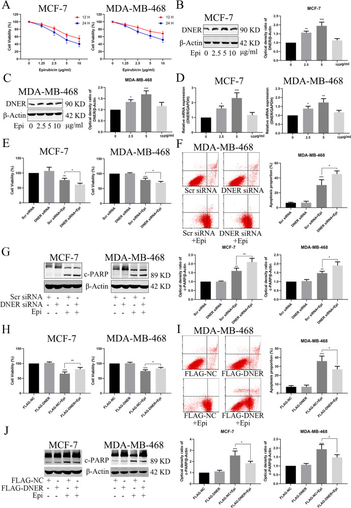
Fig. 7. DNER reduces the chemosensitivity of BC cells to epirubicin in vitro.
a Cell proliferation was detected by CCK-8 after treated with different concentrations of epirubicin in two BC cell lines. b, c DNER was analyzed by western blotting in BC cells treated as described above. Right: quantitative analysis of the optical density ratio of DNER compared with β-actin are shown. d Expression of epirubicin-induced DNER was detected by PCR. e Cell viability was assessed by CCK-8 after DNER knockdown treated with epirubicin or not. f Analysis of apoptosis with FACS in MDA-MB-468 cells treated as described in (e). Right: Quantitative analysis of apoptosis ratio. g The expression of PARP was detected by western blotting in BC cells treated as described above. Right: quantitative analysis of the optical density ratio of c-PARP compared with β-actin are shown. h Cell growth was measured by CCK-8 after DNER overexpression treated with epirubicin or not. i Analysis of apoptosis with FACS in MDA-MB-468 cells treated as described in (h). Right: Quantitative analysis of apoptosis ratio. j The expression of PARP was detected by western blotting in BC cells treated as described above. Right: quantitative analysis of the optical density ratio of c-PARP compared with β-actin are shown. The values are the mean ± SD from three independent experiments. *p < 0.05, **p < 0.01, ***p < 0.001 vs the corresponding group.

