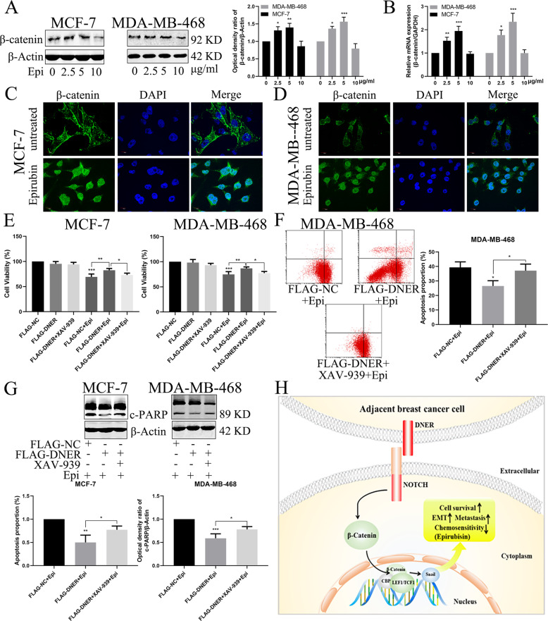
Fig. 8. Regulation of the Wnt/β-catenin pathway by DNER is involved in epirubicin-induced apoptosis.
a β-catenin was analyzed by western blotting in BC cells treated with different concentrations of epirubicin. Right: quantitative analysis of the optical density ratio of β-catenin compared with β-actin are shown. b Expression of epirubicin-induced DNER detected by real-time RT-PCR. c, d Distribution of β-catenin in BC cells treated with epirubicin that were analyzed with confocal microscopy. β-catenin is stained green, and the nucleus is stained blue. Scale bar = 10 μm. e Cells were transfected with FLAG-NC or FLAG-DNER, and then treated with XAV-939 for 24 h before epirubicin treatment. Cell proliferation was detected by CCK-8. f Analysis of apoptosis with FACS in MDA-MB-468 cells treated as described above. Right: Quantitative analysis of apoptosis ratio. g The expression of PARP was detected by western blotting in BC cells treated as described above. Right: quantitative analysis of the optical density ratio of c-PARP compared with β-actin are shown. The values are the mean ± SD from three independent experiments. *p < 0.05, **p < 0.01, ***p < 0.001 vs the corresponding group. h Schematic model for DNER-induced biological function of BC cells.

