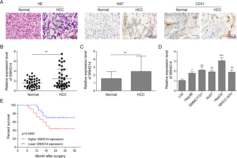
Fig. 1. SNHG14 is highly expressed in HCC tissues and cells.
a H&E staining and IHC analysis of Ki67 and CD31 in HCC and paired adjacent normal tissues. Scale bar, 50 μm. b, c qRT-PCR analysis was performed to determine the expression of SNHG14 in 40 paired HCC tissues and adjacent nontumor tissues. d The expression level of SNHG14 in L02 and different HCC cell lines were determined by qRT-PCR. GAPDH served as an internal control. e Kaplan–Meier survival curves for HCC patients with high SNHG14 expression (n = 22) and those with low SNHG14 expression (n = 18). Data were representative images or were expressed as the mean ± SD of n = 3 experiments. *P < 0.05, **P < 0.01, ***P < 0.001.

