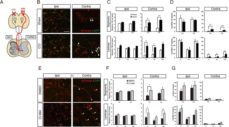
Fig. 3. The number of synapses in CST fibers is increased in the denervated side of the cervical spinal cord by HDAC inhibitor-treatment.
a Schematic illustration of the cortical injury model in this study. Cortical injury to the motor cortex (red) damages the CST (dotted blue lines). AAV-phSyn1-tdTomato-T2A-SypEGFP was injected into the contralesional (uninjured) motor cortex to label the intact CST and synapses. DMSO or CI-994 was injected once a day from 28 days after the injury. The sprouting axons (red line) of the intact CST cross the midline into the denervated side of the spinal cord (Contra) and form synapses, labeled by GFP (green). b Representative images showing AAV-labeled CST axons (red) and synaptic boutons (green) in the cervical spinal cord of Sham and CCI mice at day 42 day after brain injury. Arrowheads show synaptophysin-GFP-positive synaptic boutons. Scale bar: 20 μm. c, d Quantitative analysis of the number of GFP-labeled synapses (c) and tdTomato-labeled CST fibers (d) in the Ipsi (AAV-injected CST path thorough) or Contra (AAV-uninjected CST path through) side of the cervical cord of the Sham or CCI mice. n = 7 (Sham group), 6 (CCI group). *p < 0.05, **p < 0.01, ***p < 0.005; Student′s t test. e Representative images showing AAV-labeled CST axons (red) and synaptic boutons (green) in the cervical spinal cord of vehicle-treated (DMSO) and HDAC inhibitor-treated (CI-994) mice 42 days after brain injury. Arrowheads show synaptophysin-GFP-positive synaptic boutons. Scale bar: 20 μm. f, g Quantitative analysis of the number of GFP-labeled synapses (f) and tdTomato-labeled CST fibers (g) in the Ipsi (AAV-injected CST path thorough) or Contra (AAV-uninjected CST path through) side of the cervical cord of the vehicle-treated (DMSO) and HDAC inhibitor-treated (CI-994) mice. n = 5 (DMSO, CI-994). *p < 0.05, **p < 0.01; Student′s t test.

