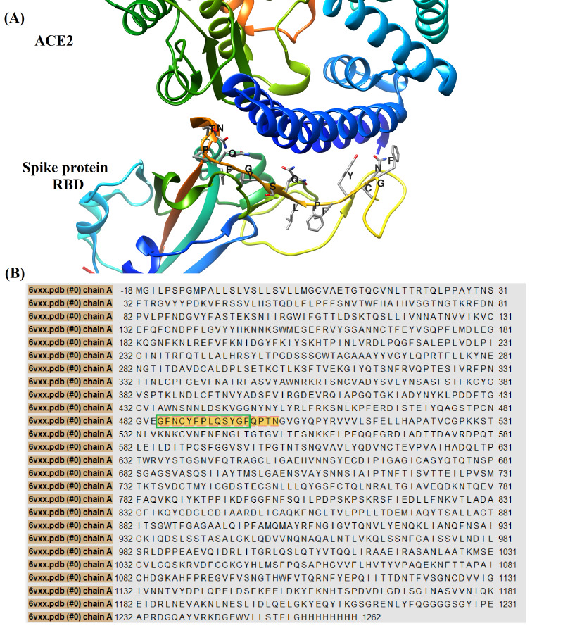
Figure 4.
(A) A three-dimensional cartoon representation for interaction between receptor binding domain (RBD) of SARS-CoV-2 spike protein and angiotensin converting enzyme 2 (ACE2). The amino acid residues of spike protein RBD involved in interaction with ACE2 were labeled in one letter format, the sequence of these residues is (GFNCYFPLQSYGFQPTN). This figure was generated by using 6M0J crystal downloaded from protein data bank [41]. We have used UCSF chimera version 1.13.1 to process and render this image [26].(B) The amino acids sequence map for chain A of SARS-CoV-2 spike protein. The residues of spike protein involved in interaction with ACE2 were colored by gold within chain A sequence, the linear B-cells epitope with sequence (GFNCYFPLQSYGF) was encircled by green line. This sequence map was generated by using PDB crystal with code (6VXX) [16,18] and UCSF chimera version 1.13.1 [26].

