
Figure 3.
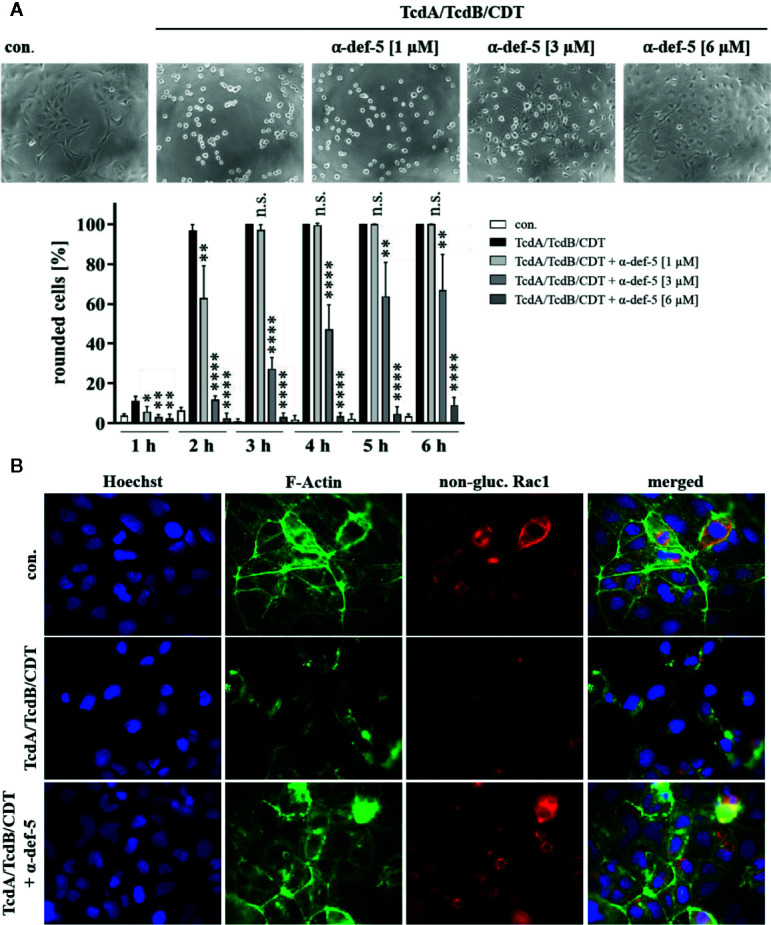
α-Defensin-5 protects cells in a time- and concentration-dependent manner from intoxication with the combination of TcdA, TcdB, and CDT. (A) Vero cells were treated with the combination of all three C. difficile toxins (TcdA: 10 pM, TcdB: 10 pM, CDT: 1 nM/1. 3 nM) and increasing concentrations of α-defensin-5 (1, 3, 6 µM). Representative images after 6 h are shown (upper panel). The amount of rounded cells was determined over time (lower panel). Values are given as mean ± SD (n=3). Significance was determined using the one-way ANOVA test (n.s. = not significant, *p < 0.05, **p < 0.01, ****p < 0.0001). (B) Caco-2 cells were treated with the combination of TcdA (10 pM), TcdB (10 pM), and CDT (2 nM/2.7 nM) in the presence or absence of α-defensin-5 (6 µM). After 5.5 h, cells were fixed, permeabilized and non-glucosylated Rac1 was stained using a specific antibody. Phalloidin-FITC was used to stain F-actin, Hoechst33342 was used to stain nuclei.