Figure 7.
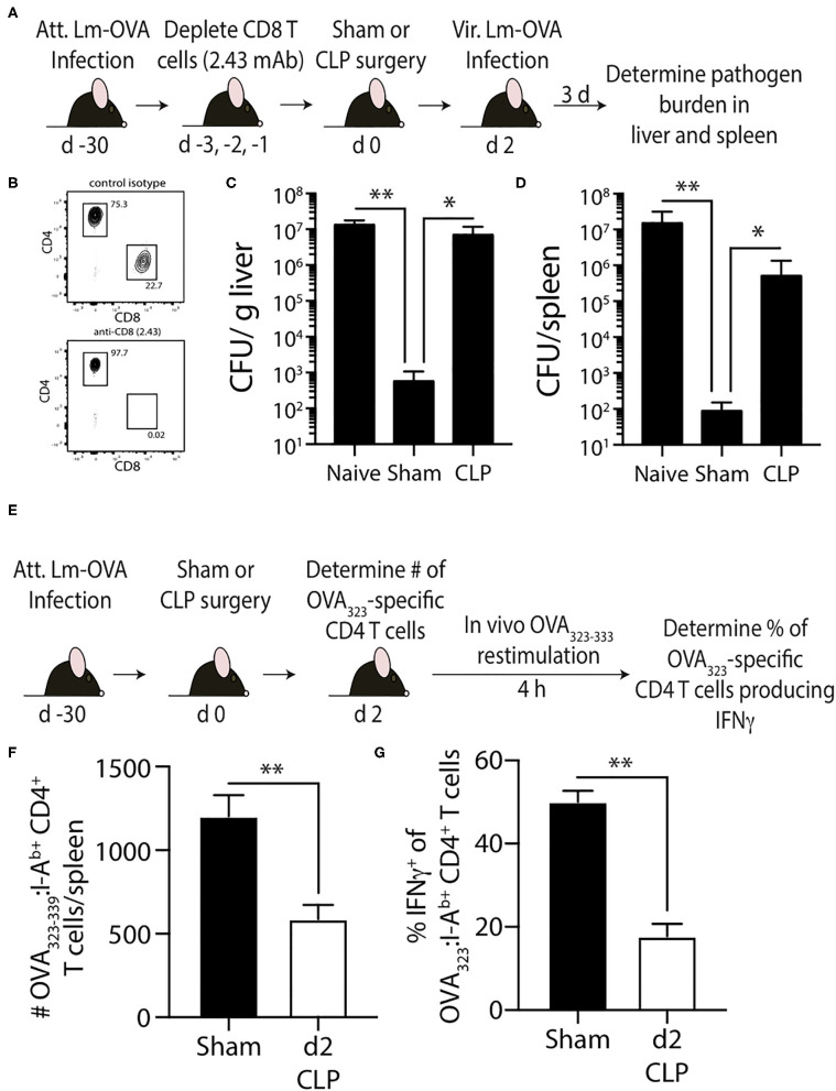
Effect of sepsis on OVA323−339-specific memory CD4 T cells. (A) Experimental design—B6 mice were infected with attenuated L. monocytogenes-OVA (LM-OVA; 107 CFU i.v.) 30 d before sham or CLP surgery. Mice in the naïve, sham, and CLP groups were depleted of CD8 T cells by injecting 100 μg anti-CD8 mAb (clone 2.43) i.v. 3, 2, and 1 days prior to surgery. (B) A small amount of blood was collected from the anti-CD8 mAb-treated mice on the day of surgery and staining for CD4 and CD8 T cells. Representative flow plots show the extent of CD8 T cell depletion compared to a reference mouse injected with a control isotype mAb. (C,D) The mice were infected with virulent LM-OVA (104 CFU i.v.) 2 days after surgery. Bacterial titers in the liver and spleen were determined 3 days post-vir LM-OVA infection. Data shown are representative of 2 independent experiments, with at least 5 mice/group in each experiment. *p ≤ 0.05 and **p < 0.01. (E) Experimental design—B6 mice were infected with attenuated L. monocytogenes-OVA (LM-OVA; 107 CFU i.v.) 30 d before sham or CLP surgery. (F) On day 2 post-surgery, the number of OVA323−339-specific memory CD4 T cells in the spleen were determined. (G) A separate cohort of mice were injected with OVA323−339 peptide (100 μg i.v.) 2 days after surgery to restimulate the OVA323−339-specific memory CD4 T cells. Spleens were harvested 4 h later, and the frequency of IFNγ+ OVA323−339-specific CD4 T cells was determined by flow cytometry. Data shown are representative of 2 independent experiments, with at least 5 mice/group in each experiment. **p < 0.01.

