
Fig 3.
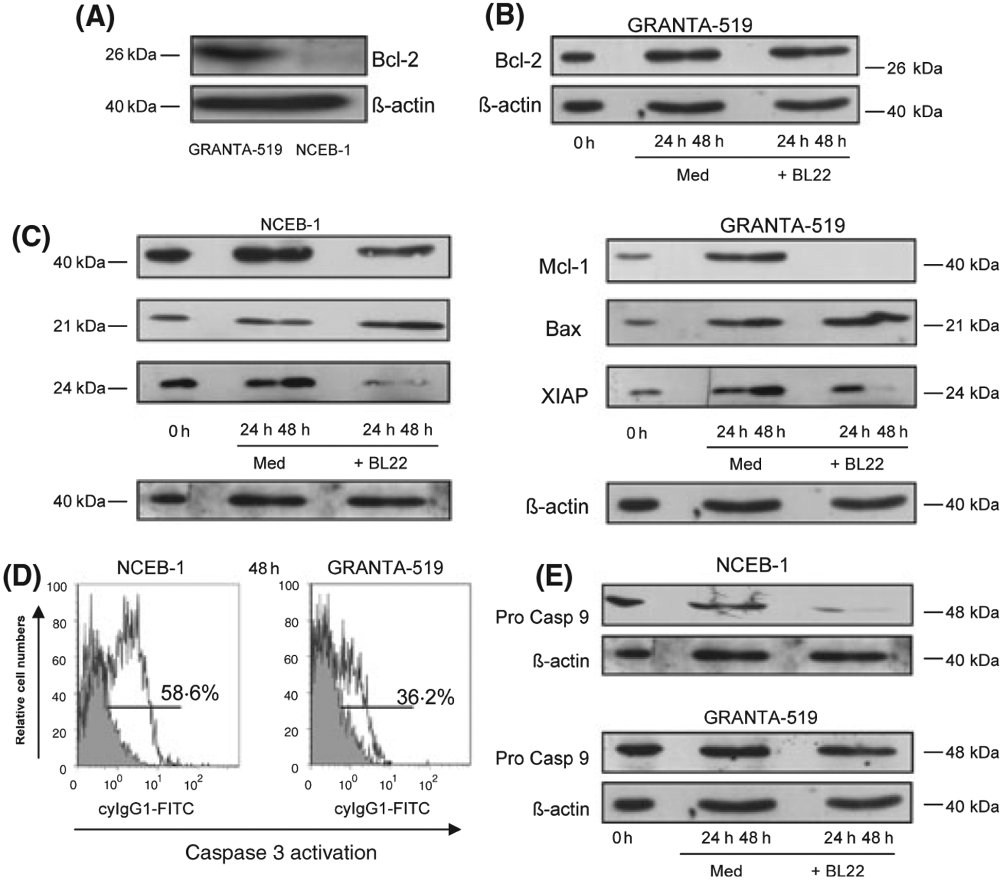
(A) Representative Bcl-2 immunoblot of GRANTA-519 cells and NCEB-1 cells. (B) Coincubation with BL22 for 24–48 h did not change Bcl-2 levels in Granta-519 cells compared to medium control, shown as a representative result. (C) Mcl-1, bax and XIAP immunoblots of NCEB-1- and GRANTA-519 cells were performed in medium (control) and together with BL22 in a concentration of 100 ng/ml for 24–48 h. (D) Representative results of flow cytometric analysis of caspase 3 activation in NCEB-1 and GRANTA-519 cells after incubation with BL22 at a concentration of 100 ng/ml for 48 h and (E) immunoblot results of procaspase 9 after BL22 incubation for the indicated time points. Anti β-actin blotting was used as loading control.