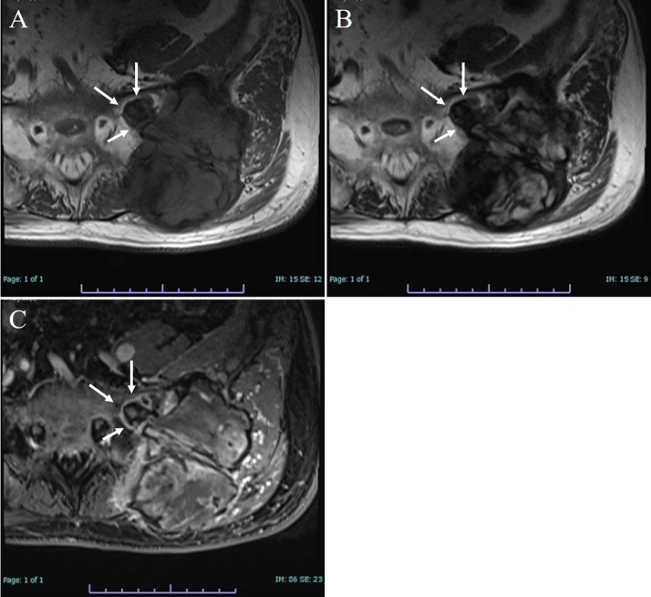
Fig. 3.
Representative MRI images. The tumor is localized to the left iliac bone involving the gluteus maximus, gluteus medius, paravertebral muscle, and sacral bone through the sacroiliac joint (white arrows). (A) T1-weighted image exhibits iso~low-signal intensity. (B) T2-weighted image exhibits inhomogeneous high signal intensity within low signal intensity. (C) Enhanced MRI exhibits inhomogeneous enhancement. MRI, magnetic resonance imaging.

