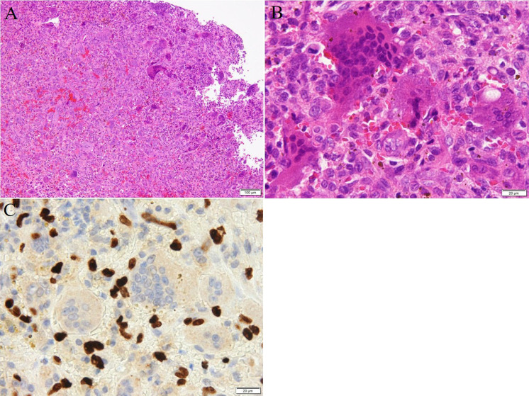
Fig. 4.
Histology and immunohistochemistry of the tumor. Staining methods include (A), (B), hematoxylin and eosin, and (C), immunohistochemistry using an antibody against histone 3.3 G34W. (A) Oval-shaped mononuclear cells are admixed within a large number of osteoclast-like giant cells (scale bar: 100 μm). (B) At high-power magnification, the giant cells having a variable number of nuclei (up to 50) and abundant eosinophilic cytoplasm are observed (scale bar: 20 μm). They are not atypical, and the marked nuclear pleomorphism and hyperchromasia are not observed. (C) A positive signal for histone 3.3 G34W in the mononuclear cells.

