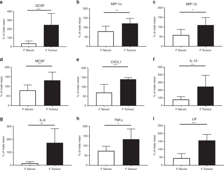
Fig. 4. Female mice have steeper cytokine gradients between periphery and the TME.
Females show significant difference in the relative quantity of cytokines in the serum and organ culture as compared to males. These values tend to be reduced compared to males in the serum but increased in the TME. The pattern is seen in chemotactic cytokines a GCSF, b MIP-1α, c MIP-1β, d MCSF and e CXCL1, as well as in f IL-10, g IL-6, h TNF-α and i LIF. Values are expressed as a percent of the cytokines from female compared to the male mean in the same location (*p < 0.050, **p < 0.010, ***p < 0.001). n = 7–9.

