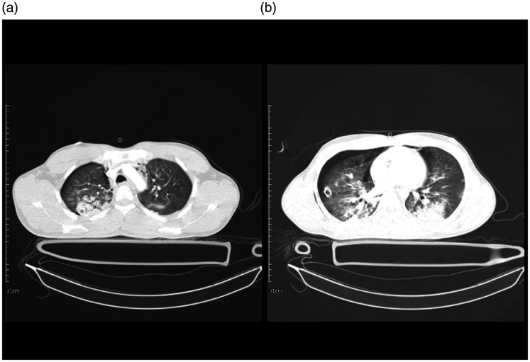
Figure 6.
A 19-year-old man with multiple trauma and lung injury and a case of COVID-19. 6 A: an axial image of the chest CT scan in the level of aortic arch showed mild pneumothorax on the right side. There are multiple round shape GGO and consolidation in both upper lobes with right-side predominance. Also, minimal pneumomediastinum is noted. 6B: evidence of pneumomediastinum and right-sided pneumothorax with a chest tube is seen. Also, consolidation in the posterior aspect of both lungs associated with faint GGO is seen.

