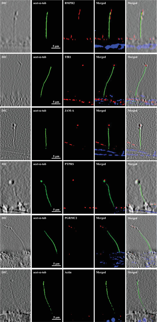
Figure 2.

Selected cELV proteins are expressed in subdomain of primary cilia. Selected proteins (red) were localized to cELV. Acetylated α‐tubulin (green) and DAPI (blue) were used as ciliary and nuclear markers. High‐resolution differential interference contrast (DIC) images confirmed the presence of cilia. Actin was used as a negative control. Ciliary marker acetylated‐α‐tubulin = acet‐α‐tubulin.
