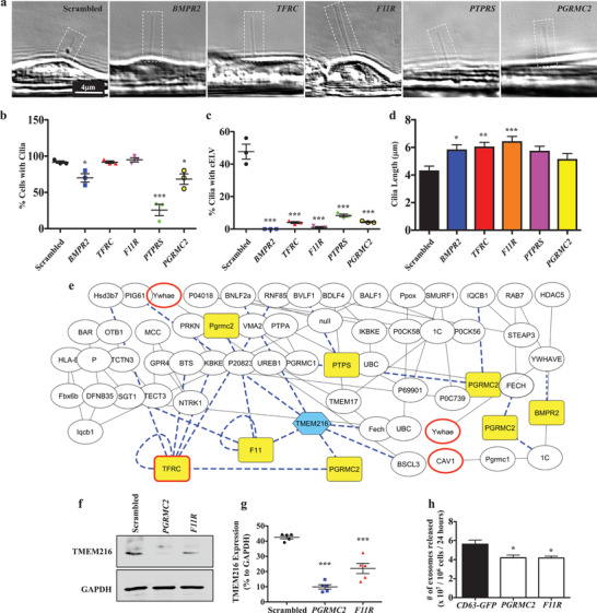
Figure 3.

Selected cELV proteins are involved in cELV formation and suppression of TMEM216 expression. a) A representative DIC images showing all corresponding knockdown genes impact on cilia and/or cELV formation. b,c) The effects of cELV proteins on cilia formation and cELV formation were quantified in BMPR2, TFRC, F11R, PTPRS and PGRMC2 knockdown cells. d) The averaged cilium length is shown in the bar graph. The effect of the knockdown on ciliary length distributions is shown in the histogram (Figure S6d, Supporting Information). N = 3 with 40 measurements for each N. For the cELV measurements, total cELV from replicates are shown because most cilia did not have cELV. e) A network of protein–protein interactions analyzed from the proteomic studies shows the functional interface of the five selected cELV proteins (highlighted in yellow) with TMEM216 (highlighted in blue) and known EV marker proteins (bolded red boarder). Dotted blue lines represent an interaction with the selected ciliary proteins or TMEM216. f,g) A quantitative Western blot analysis of TMEM216 expression was performed in PGRMC2 or F11R knockdown cells. N = 5 for Western blot. h) The cELV secretion was quantified in PGRMC2 or F11R knockdown cells. N = 3 for cELV quantification. *, p < 0.05; **, p < 0.01; ***, p < 0.001; compared with the scrambled control.
