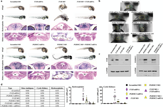
Figure 4.

Repressed cELV protein expression results in ciliopathy phenotypes, pericardiac edema, and randomized heart position in zebrafish. a) Fish were injected with scrambled, specific F11R or PGRMC2 mRNA, morpholino (MO), and both mRNA plus morpholino. Representative phase contrast images show zebrafish at 48 and 72 h postfertilization (hpf). The H&E images represent fish at 72‐hpf; black and red asterisks indicate hydrocephalus and renal cysts, respectively. nc = notochord. b) In addition to pericardiac edema (Figure S12, Supporting Information), randomized heart looping was observed in PGRMC2 MO fish (Movie S9, Supporting Information) Yellow‐ and green‐dotted circles show ventricle and bulbus arteriosus, respectively. c) Confirmation of the rescue phenotype was confirmed with the corresponding protein expression via Western blot analysis. d) A quantitative analysis of asymmetry, kidney cysts and hydrocephalus depict the frequency and severity of the phenotypes. N = 40–60 fish per group. *, p < 0.05; **, p < 0.01; compared with the scrambled control.
