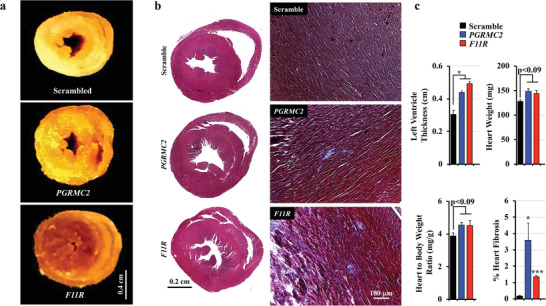
Figure 8.

Mice lacking of cELVs have left ventricular hypertrophy and cardiac fibrosis. a) The thickness of the left ventricle was compared in transverse cut of whole mount hearts, showing left ventricular hypertrophy in PGRMC2 and F11R mice. b) Representative images of H&E and Masson's Trichrome staining show left and right ventricles with significant fibrosis in the left ventricular endocardial regions of hearts from the PGRMC2 and F11R knockdown mice. c) Left ventricular thickness, heart weight, heart‐to‐body weight ratio and percent of fibrosis in the heart were quantified. N = 3 mice in each scrambled control, PGRMC2 and F11R knockdown groups. *, p < 0.05; ***, p < 0.001; compared with the corresponding scrambled control group.
