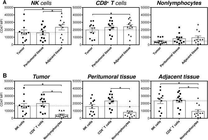
Figure 3.
Intensities of CD47 staining of NK cells, CD8+ T cells, and nonlymphocytes in the tumoral and paratumoral compartments of EC patients. (A) The analyzed cells were gated as in Fig. 1A, and the mean fluorescence intensities of CD47 staining (CD47 MFI) of NK cells (CD45+CD3−CD56+), CD8+ T cells (CD45+CD3+CD8+), and nonlymphocytes (CD45−) in tumor tissue, peritumoral tissue and adjacent tissue samples from 12 EC patients were determined by flow cytometry. (B) The CD47 MFIs determined in (A) were evaluated within each tissue compartment. In (A) and (B), the significance of differences was determined by nonparametric one-way ANOVA with Dunn's multiple comparison test (n = 12). *P < 0.05 was considered significant. The data are shown as the mean ± SEM.

