Figure 1.
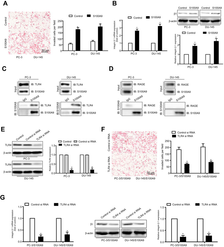
S100A9 promotes prostate cancer cell invasion and β1 integrin expression through interaction with TLR4. PC-3 and DU-145 cells were treated with S100A9 (20 µg/ml) for 48 h. (A) PC-3 and DU-145 cells invasion was measured by transwell invasion assay. (B) The mRNA and protein levels of β1 integrin were determined by qPCR or Western blot. PC-3 and DU-145 cells were treated with S100A9 (20 µg/ml) for 48 h. (C, D) Cell extracts were immunoprecipitated (IP) with control mouse IgG, mouse anti-S100A9 antibody. Immunoblot (IB) was used to detect S100A9, TLR4 and RAGE. PC-3 and DU-145 cells were transfected with TLR4 or control siRNA. (E) TLR4 expression was examined by Western blot after 48 h siRNA transfection. PC-3 and DU-145 cells were transfected with TLR4 or control siRNA followed by stimulation with S100A9. (F) Tumor cell invasion was measured by transwell invasion assay. (G) The mRNA and protein levels of β1 integrin were determined by qPCR or Western blot. Scale bar 50 μ. Magnifcation×200. Data are represented as the mean ± S.E.M. *p<0.05.

