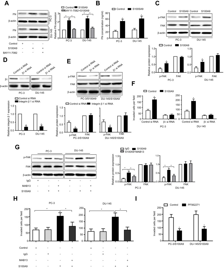
Figure 3.
S100A9 promotes prostate cancer cell invasion via integrin β1/FAK signaling. PC-3 and DU-145 cells were treated with or without BAY11-7082 (5µM) for 30 min. Then cells were treated with or without S100A9 (20 µg/ml) for 48 h. (A) Fibronectin expression was determined by Western blot. PC-3 and DU-145 cells were treated with or without S100A9 (20 µg/ml) for 48 h. (B) Supernatant fibronectin (FN) concentration was determined by ELISA. PC-3 and DU-145 cells were treated with or without S100A9 (20 µg/ml) for 30 min. (C) The phosphorylation of FAK was measured by Western blot. PC-3 and DU-145 cells were transfected with control siRNA or integrin β1-specific siRNA for 48 h. Then cells were treated with or without S100A9 (20 µg/ml) for 30 min, the expression of integrin β1 (D) or phosphorylation of FAK (E) was measured by Western blot. PC-3 and DU-145 cells were transfected with control siRNA or integrin β1-specific siRNA for 48 h. Then cells were treated with or without S100A9 (20 µg/ml) for 48 h. (F) The invasion activity were measured by transwell invasion assay. (G) PC-3 and DU-145 cells were treated with β1 integrin functional blocking antibody MAB13 (50 μg/mL) or control IgG (50 μg/mL) for 30 min and then treated with or without S100A9 (20 µg/ml) for 30 min and the phosphorylation of FAK was measured by Western blot. (H) PC-3 and DU-145 cells were treated with MAB13 (50 μg/mL) or control IgG (50 μg/mL) for 30 min and then treated with or without S100A9 (20 µg/ml) for 48 h for invasion. (I) PC-3 and DU-145 cells were pretreated for 30 min with FAK inhibitor, PF562271 (100 nM) followed by stimulation with S100A9 (20 µg/ml) for 48 h for invasion. Data are represented as the mean ± S.E.M. *p<0.05.

