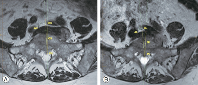
Fig. 4.

Reduction in size of bare window area of L5/S1 disk by position, with left common iliac vein crossing over the midline. (A) Supine position MRI and (B) lateral decubitus MRI. ASBS & ALBL, L5/S1 bare window; OSPSBS & OLPBL’, oblique angle; AS & AL, right common iliac vein; BS & BL, left common iliac vein; OS & OL, center of L5/S1 disk; PS & PL, posterior disc midpoint; MRI, magnetic resonance imaging.
