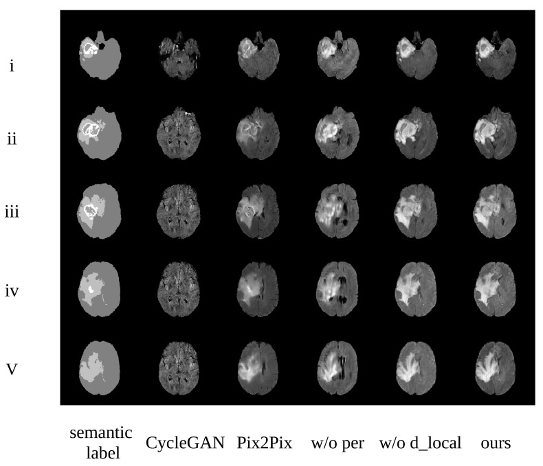
Figure 8.
Examples generated from different methods. i–v represent slices 50, 60, 70, 80, and 90 from this patient, respectively. Images from the left to the right are the corresponding semantic label, CycleGAN, Pix2Pix, TumorGAN without regional perceptual loss, TumorGAN without the local discriminator, and TumorGAN.

